

Introduction
Tube shunt surgery is a crucial intervention for managing glaucoma, especially for patients who do not respond well to conventional treatments. This step-by-step tutorial delves into the intricacies of the surgery, offering insights into its purpose, procedural techniques, and post-operative care. Patients often find it difficult to navigate the complexities of treatment options and potential complications. By mastering these complexities, patients can significantly improve their treatment outcomes in the fight against glaucoma.
Define Tube Shunt Surgery and Its Role in Glaucoma Treatment
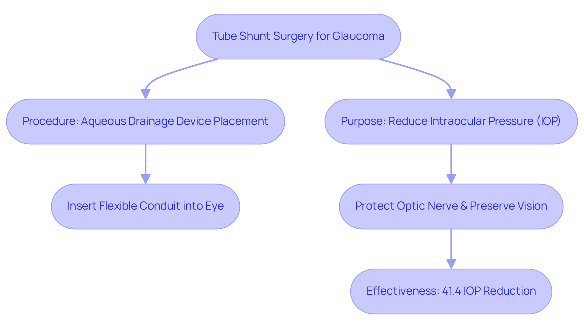
Tube shunt surgery glaucoma, also referred to as aqueous drainage device placement, serves as a critical intervention for patients struggling to manage their glaucoma effectively. This procedure, known as tube shunt surgery glaucoma, is recommended for those who often struggle to manage their glaucoma effectively with standard treatments, making it a necessary option.
During the procedure, a small, flexible conduit is inserted into the eye, connecting to a silicone drainage pouch that aids in the outflow of excess fluid, thereby reducing intraocular pressure (IOP). By effectively controlling IOP, tube shunt surgery glaucoma is intended to protect the optic nerve and preserve vision in patients with advanced glaucoma.
Recent research shows that drainage implant procedures have a cumulative failure probability of 29.8% at five years, compared to 46.9% for trabeculectomy, emphasizing its effectiveness in long-term glaucoma treatment. Furthermore, the procedure has demonstrated a notable decrease in IOP, with an average reduction of 41.4% observed in individuals undergoing this treatment, while trabeculectomy resulted in a 49.5% reduction in IOP over five years.
These results highlight the significance of drainage device procedures as a feasible choice for individuals encountering difficulties in managing their glaucoma, especially since insufficient IOP reduction is the leading reason for treatment failure. Given the high failure rates of traditional treatments, tube shunt surgery glaucoma represents a promising option for patients with advanced glaucoma.
Explain the Surgical Procedure: Step-by-Step Guide to Tube Shunt Surgery
Tube Shunt Surgery Procedure
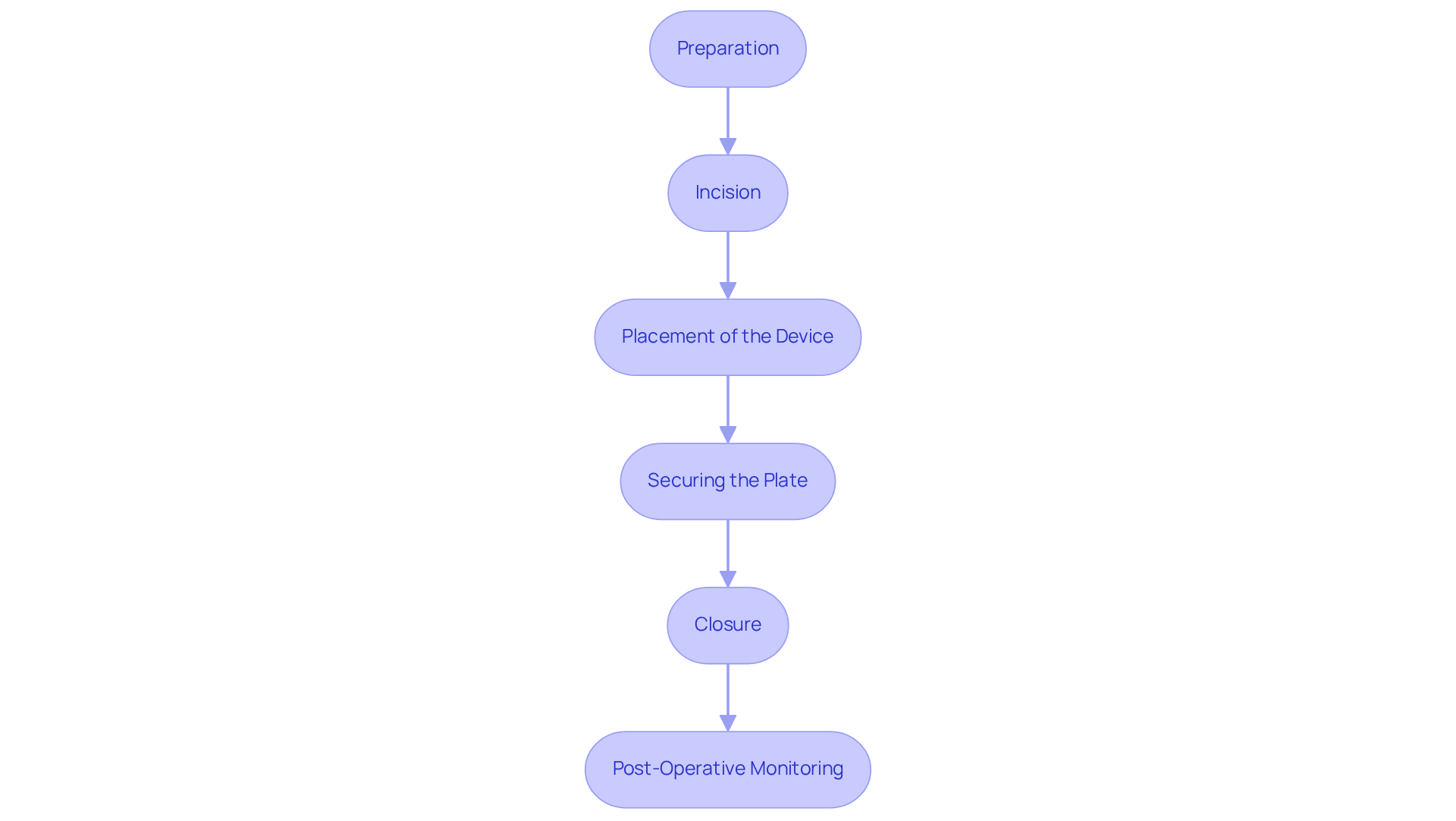
- Preparation
Tube shunt surgery is a critical procedure aimed at managing intraocular pressure in patients with glaucoma. The patient is comfortably positioned, and local anesthesia is administered to numb the eye. The team may provide sedation to ensure the patient remains relaxed throughout the procedure. Typically, the surgery lasts between 45 to 60 minutes. - Incision
A small cut is made in the conjunctiva, the thin membrane covering the eye, allowing access to the area where the device will be placed. - Placement of the Device
A small, flexible instrument is inserted into the anterior chamber of the eye, positioned just in front of the iris. This conduit connects to a drainage plate that will be positioned beneath the conjunctiva. - Securing the Plate
The drainage plate is secured beneath the conjunctiva to ensure it remains in place and functions effectively, facilitating proper drainage of aqueous humor. - Closure
The surgeon sutures the conjunctiva closed and patches the eye to protect it during the crucial initial healing phase. - Post-Operative Monitoring
After the procedure, the patient is monitored for any immediate complications, such as elevated eye pressure or inflammation, before being discharged, typically within a few hours. Follow-up visits are essential to ensure the drainage device is operating properly and to address any possible complications. Effective post-operative care, including adherence to prescribed medications and regular check-ups, is essential for optimal recovery following tube shunt surgery glaucoma. Without proper post-operative care, the risk of complications increases significantly, potentially jeopardizing the success of the surgery.
Discuss Post-Operative Care and Complications of Tube Shunt Surgery
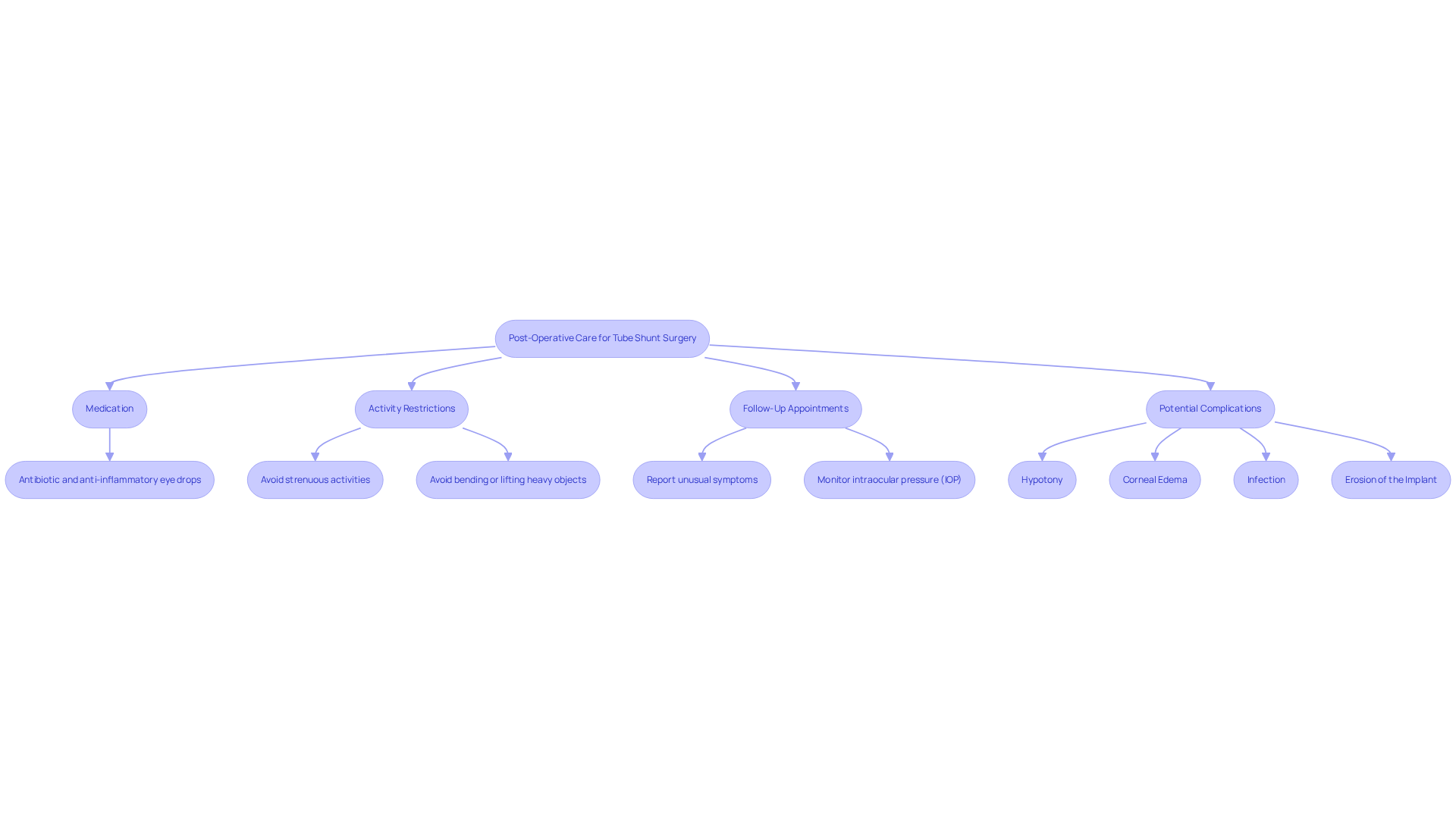
Post-operative care following tube shunt surgery is critical for ensuring optimal recovery and minimizing complications:
-
Medication: Patients are typically prescribed antibiotic and anti-inflammatory eye drops to prevent infection and reduce inflammation.
-
Activity Restrictions: Avoid strenuous activities, bending, or lifting heavy objects for several weeks to prevent pressure on the eye. Lifting more than 10 pounds should be strictly avoided during recovery.
-
Follow-Up Appointments: Regular follow-up visits are essential to monitor intraocular pressure (IOP) and assess the healing process. Patients should report any unusual symptoms, such as increased pain or vision changes, to their eye care team promptly.
-
Potential Complications: While tube shunt surgery is generally safe, complications can occur, including:
- Hypotony: Low eye pressure that can lead to blurred vision, with a reported incidence of hypotony-related complications.
- Corneal Edema: Swelling of the cornea due to contact with the device, which may require monitoring.
- Infection: As with any medical procedure, there is a risk of infection, necessitating vigilance in post-operative care.
- Erosion of the Implant: The device may deteriorate through the conjunctiva, necessitating additional intervention, with an overall incidence of exposure at 2.0%, which can result in complications if not addressed promptly.
Adhering to these post-operative care guidelines is essential for maximizing recovery and ensuring long-term success. Data indicates that over 20% of eyes that undergo drainage device procedures necessitate further interventions, underscoring the importance of diligent post-operative care.
Highlight Technological Advances in Tube Shunt Surgery
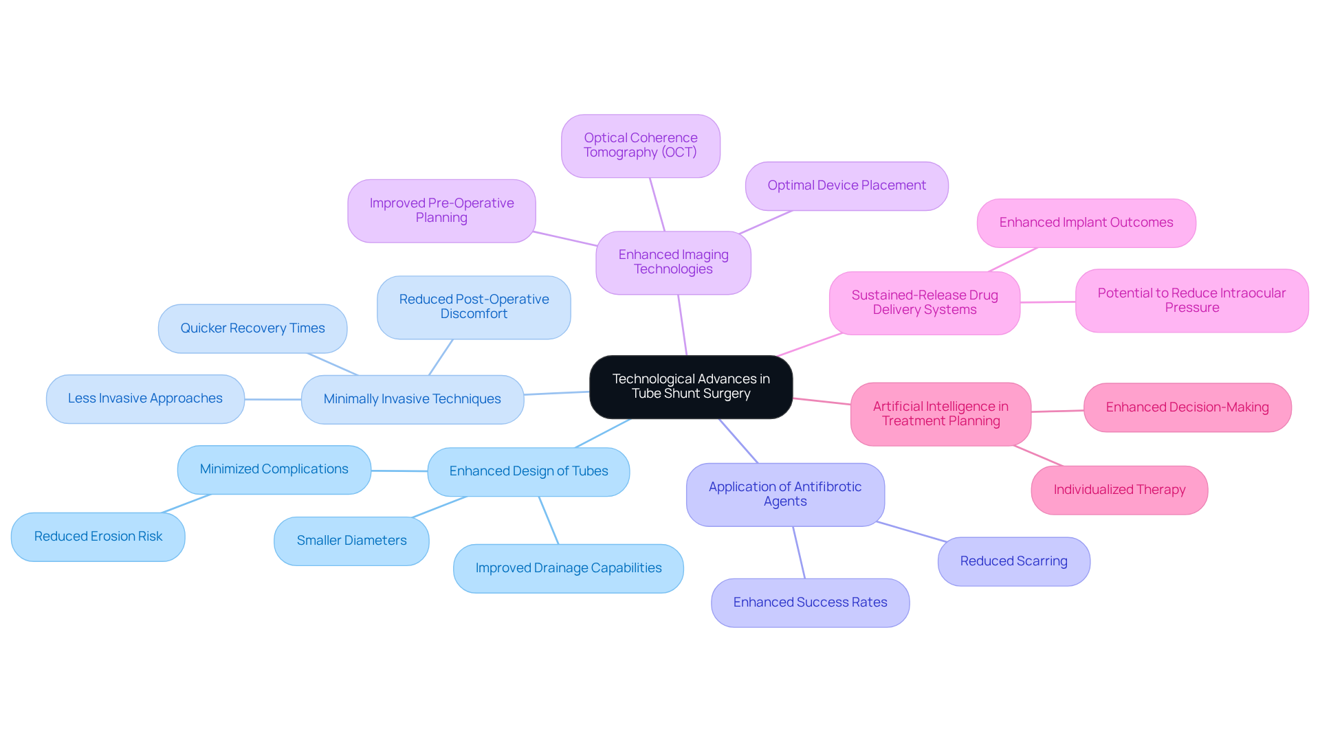
Recent advancements in drainage device procedures have transformed patient outcomes, yet challenges remain in minimizing complications. Key innovations include:
- Enhanced Design of Tubes: Newer drainage devices, such as the Ahmed ClearPath, feature smaller diameters and improved drainage capabilities, which assist in minimizing complications like erosion.
- Minimally Invasive Techniques: Advances in surgical methods allow for less invasive approaches, leading to quicker recovery times and reduced post-operative discomfort.
- Application of Antifibrotic Agents: The supplementary application of antifibrotic agents during procedures has been demonstrated to enhance the success rates of implanting drainage devices by reducing scarring around the implant.
- Enhanced Imaging Technologies: The integration of advanced imaging technologies, such as Optical Coherence Tomography (OCT), facilitates improved pre-operative planning and post-operative monitoring, ensuring optimal placement and function of the device.
- Sustained-Release Drug Delivery Systems: Emerging technologies, including sustained-release drug delivery systems, are being researched for their potential to reduce intraocular pressure, which could enhance outcomes of implant procedures.
- Artificial Intelligence in Treatment Planning: AI is being investigated for its capability to enhance treatment planning in glaucoma, potentially improving the decision-making process for drainage procedures.
These advancements not only make tube shunt surgery more effective but also significantly enhance the patient experience. As these innovations continue to evolve, they promise to redefine the standards of care in glaucoma management, ultimately enhancing patient outcomes.
Conclusion
Tube shunt surgery is a critical option for patients with glaucoma who find traditional treatments inadequate. This procedure helps control intraocular pressure and is vital for preserving vision in patients with advanced glaucoma. Tube shunt surgery uses a small conduit to drain fluid, significantly lowering the risk of optic nerve damage, making it an important choice in glaucoma management.
Throughout the article, we have explored key aspects of tube shunt surgery, including:
- A detailed step-by-step guide to the surgical procedure
- Essential post-operative care
- Potential complications
Innovations in technology, such as enhanced drainage device designs and minimally invasive techniques, have further improved patient outcomes and reduced recovery times. Staying updated on these advancements is crucial for ensuring the best treatment options for glaucoma.
As the field of glaucoma management continues to evolve, it is essential for patients in the North Texas region, including those in Plano and Las Colinas, to consider tube shunt surgery as a viable option. For those interested in exploring this procedure or seeking expert guidance, scheduling a consultation with Brooks Eye Associates can provide valuable insights and personalized care. Considering tube shunt surgery could be the key to maintaining vision and improving life quality for those affected by glaucoma.
Frequently Asked Questions
What is tube shunt surgery and its purpose in glaucoma treatment?
Tube shunt surgery, also known as aqueous drainage device placement, is a procedure designed for patients who struggle to manage their glaucoma effectively with standard treatments. It aims to reduce intraocular pressure (IOP) to protect the optic nerve and preserve vision in individuals with advanced glaucoma.
How does tube shunt surgery work?
During tube shunt surgery, a small, flexible conduit is inserted into the eye, connecting to a silicone drainage pouch. This setup aids in the outflow of excess fluid, thereby reducing IOP.
What are the success rates of tube shunt surgery compared to other treatments?
Recent research indicates that tube shunt surgery has a cumulative failure probability of 29.8% at five years, which is lower than the 46.9% failure rate for trabeculectomy. Additionally, tube shunt surgery has shown an average IOP reduction of 41.4%, while trabeculectomy resulted in a 49.5% reduction over the same period.
Why is tube shunt surgery considered a viable option for glaucoma patients?
Tube shunt surgery is considered a promising option for patients with advanced glaucoma, especially since insufficient IOP reduction is the leading cause of treatment failure. Its effectiveness in long-term management makes it a critical intervention for those facing challenges with traditional treatments.