
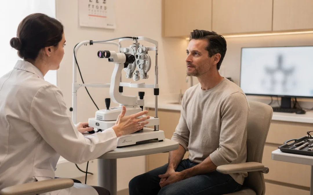
Read the 20/20 number as a measurement of acuity
Visual acuity sharper than 20/20 describes clarity measured on an eye chart. Visual acuity measures the clearness or sharpness of vision, not every way your eyes work. A 20/20 result means you can see at 20 feet what a person with normal sight can see at 20 feet on the chart.
The U.S. visual-acuity fraction has a specific meaning. The top number represents your distance from the chart, while the bottom number identifies the last line you read correctly. A larger bottom number represents lower visual acuity: with 20/30 vision, you can read at 20 feet what normal sight can read at 30 feet.
A 20/15 result is sharper than average. With 20/15 vision, you can read a line at 20 feet that average sight can read only at 15 feet. That result describes chart acuity under the conditions of the test; it does not establish that your eyes are completely healthy or that every visual task will feel effortless.
A Snellen eye chart is commonly used to test visual acuity at 20 feet. A Random E test can assess acuity without relying on familiar letters, and simplified symbol or letter testing can be used for children. The test answers a narrow question: how clearly you can identify the displayed target at a defined distance. Near-vision charts answer a different question from distance-vision charts.
Separate sharper acuity from everyday visual quality
Visual acuity can be sharper than 20/20 without becoming a complete definition of vision. In the United States, 20/20 is commonly considered normal visual acuity, while maximum human acuity is finer at 20/16 to 20/12. A chart result describes resolution in one test; it does not measure every aspect of contrast, color, peripheral vision, or eye health.
Contrast sensitivity affects how easily you distinguish an object from its background, especially when lighting or visual detail changes. The cited pilot study associated increasing age, small pupil diameter, farsightedness, and bright-light testing conditions with lower contrast sensitivity in healthy individuals. The result applies to that study population and testing pattern, not to every visual situation.
Age, pupil size, farsightedness, and lighting conditions can affect contrast sensitivity. Aging also affects macular pigments, photoreceptors, and neural paths in the retina. Retinal changes can explain decreases in light sensitivity, contrast sensitivity, and visual acuity, as well as prolonged dark adaptation, in individuals over age 50. The overall evidence is not uniform: some studies find little appreciable decrease with age, while most report declines in photopic and scotopic contrast sensitivity. The type of contrast and the visual condition tested can change the result. Studies have noted a much larger decrease in rods than cones in the aging retina.
A 20/30 result illustrates the direction of the scale: at 20 feet, you can read letters that average sight can see from 30 feet. A result below 20/20 can reflect a refractive issue, but a result above 20/20 still does not measure every aspect of how you see in dim light, against glare, at close range, or during a visually demanding activity.

Use the symptom pattern to explain why 20/20 may not feel clear
A 20/20 visual-acuity result can coexist with color-vision differences, reduced contrast, or difficulty tracking fast-moving objects. An eye chart alone does not determine whether you have glaucoma or a retinal problem, and it does not measure loss of peripheral vision. For that reason, a clear chart result should not be treated as proof that every part of your visual system is normal.
Presbyopia can explain why distance vision looks sharp while reading becomes difficult. Presbyopia is common after age 40 and can make near tasks such as reading or sewing harder; blurry close-up vision may begin around age 40. Reading glasses, some contact lenses, and refractive surgeries can help with near-focus difficulty, but a distance acuity number does not tell you which approach fits your near-vision needs.
Digital eye strain can also make clear vision feel inconsistent. Digital eye strain affects people of all ages and might cause blurred vision, aching or uncomfortable eyes, dryness, tearing, or stinging after prolonged device use.
Refractive errors remain a common explanation for blur below 20/20. Nearsightedness, farsightedness, and astigmatism cause blurry vision in most cases described by the American Academy of Ophthalmology source. Glasses or contact lenses can be prescribed with the goal of bringing vision to 20/20, but your symptoms may involve more than refractive sharpness when you already measure 20/20.
Use your symptom pattern as part of the question you bring to an appointment:
- Distance blur suggests a question about refraction or another visual pathway.
- Reading difficulty suggests a separate near-focus question, particularly after age 40.
- Glare, reduced contrast, or trouble following moving objects calls for more than a single acuity-line result.
- Blur, dryness, stinging, or aching after prolonged screen use may point toward an ocular-surface or eye-strain discussion.
The practical boundary is simple: 20/20 tells you how you performed on that acuity test, not why every visual task feels the way it does. Before an eye appointment, record whether blur occurs at distance or near, in dim light or glare, or after prolonged screen use. A record of symptom triggers can help an ophthalmologist compare a 20/20 or sharper acuity result with the visual conditions that matter during daily activities.
Turn the goal into an evaluation question
A comprehensive examination is more accurate than a home vision test and may detect health issues or eye diseases that have no noticeable symptoms. Before refractive surgery, a comprehensive medical eye evaluation should be performed.
A refractive-surgery examination assesses more than the smallest line you can read. The assessment should include:
- Corrected and uncorrected distance and near acuity
- Refraction to measure the focusing error
- Corneal topography or tomography to evaluate corneal shape
- Corneal-thickness measurement
- Tear-film and ocular-surface evaluation
- Eye-movement and alignment assessment
These findings help separate a correctable refractive concern from a near-focus, ocular-surface, alignment, corneal, or broader eye-health question. They also give the ophthalmologist information needed before discussing whether any procedure is relevant. Corneal topography or tomography also helps assess the optical state of the cornea and is relevant for toric IOL implantation or possible keratorefractive treatment after lens surgery. A corneal-thickness measurement supplies structural information that a visual-acuity result does not provide, so a sharp chart result cannot replace the examination data used in a refractive-surgery discussion.
Prescription stability and healing factors also belong in the discussion. A prescription change in the past year indicates refractive instability, and factors that make vision fluctuate should be discussed with your doctor. Some autoimmune diseases, immunodeficiency states, diabetes, and medications may prevent proper healing after a refractive procedure. In the cited FDA guidance, LASIK is not approved for people under age 18.
A patient study reported that all three treated eyes completed the study with 20/20 vision or better. The same study reported that all but one subject completed it with at least 20/20 vision without glasses, while one subject had 20/32 vision without glasses. The three-eye result describes the study’s treated eyes, and the 20/32 result describes one subject; neither result predicts what an evaluation or procedure would produce for you.
Compare correction paths by what they change
Refractive surgery includes elective procedures that modify the eye’s refractive status. The relevant question is not simply whether an option can produce a sharper chart result; it is what part of the eye the option changes and whether your examination findings make that discussion appropriate. Refractive surgery may be considered when you want to be less dependent on eyeglasses or contact lenses, including for occupational or cosmetic reasons.
| Option | What it changes | When it may enter the discussion |
|---|---|---|
| LASIK | A laser reshapes the surface of the cornea. | It may be discussed when the goal is improved vision or less dependence on glasses, after the examination addresses corneal and ocular-surface findings. |
| PRK or another surface-ablation procedure | A corneal refractive approach that does not require creating a LASIK flap. | It might be considered when certain corneal, healing, trauma-risk, or dry-eye factors are present. |
| SMILE | A refractive procedure considered in some situations instead of creating a LASIK flap. | It might be considered when particular corneal or other examination factors make that discussion relevant. |
| Phakic lens placed in front of the natural lens | An implant is placed in front of the natural lens, which is not removed. | It is designed for some high refractive errors that cannot be safely corrected with corneal surgery. |
| Refractive lens exchange | An artificial lens replaces the eye’s natural lens. | It may enter the discussion when lens-based correction is relevant; the procedure is performed much like cataract surgery. |
Our Custom Lens Replacement is designed as an alternative to LASIK for patients over 40 or those with certain eye conditions. That description identifies a discussion path, not a determination that the option is suitable for you.
PRK, another surface-ablation procedure, or SMILE might reduce risks associated with creating a LASIK flap when certain findings are present, but those risks are not eliminated entirely. A phakic lens preserves the natural lens rather than replacing it, while refractive lens exchange replaces the natural lens; those are different surgical changes, not interchangeable labels for sharper acuity.
Follow-up and device-specific conditions require separate discussion. If further correction or enhancement is necessary, wait until eye measurements are consistent for two consecutive visits at least 3 months apart before re-operation. For EVO ICL, an FDA clinical study of Visian ICL lenses for myopia reported that some patients had a loss of 30% or more of their corneal endothelial cells; that percentage measures cell loss within affected patients, not the share of the study population. These details are reasons to discuss the specific procedure, its conditions, and its alternatives rather than choosing from a chart result alone. Contact lenses can change corneal shape for up to several weeks after discontinuation, depending on the lens type. Before a refractive-surgery evaluation, follow the doctor’s instructions about stopping lenses and repeating measurements; measurements may need to be repeated at least a week after the initial evaluation, especially for RGP or hard lenses.
Account for age and near-vision goals
Distance sharpness and near focus are separate goals. Presbyopia is common after age 40 and can make it more difficult to focus at near for activities such as reading or sewing. If you want sharper distance vision but also struggle with menus, books, phones, or close work, make both goals explicit during the evaluation.
A study of 91 subjects observed decreased contrast sensitivity at high spatial frequencies but no effect at lower frequencies in subjects over 40 years old. The 91-subject result shows why the type of visual task and contrast condition matters; it does not establish a universal age-related outcome for your vision. Aging-related changes in the retina can affect light sensitivity, contrast sensitivity, visual acuity, and dark adaptation, although studies differ in how consistently they find those effects.
Reading glasses, some contact lenses, and refractive surgeries can help with near-focus difficulty associated with presbyopia. Glasses or contacts have a goal of bringing your vision to 20/20, but a distance result sharper than 20/20 does not remove the separate question of how you will focus up close. Ask for an explanation of how each discussed option relates to distance acuity, near work, and your daily visual priorities.
Prepare three goals before the appointment:
- Describe the distance tasks you want to see more clearly.
- Describe the near tasks that remain difficult, including when the difficulty began.
- Decide how much dependence on glasses or contacts you hope to reduce, without treating a particular chart number as a promised result.
This list keeps the discussion focused on how you use your vision, not only on the smallest line you can read.
Plan the financial conversation around the procedure
LASIK typically ranges from $1,500 to $3,500 per eye in Texas, with an average cost around $2,250. This is a general reported range per eye, not a combined two-eye total or a personal estimate.
Laser eye surgery prices vary greatly according to factors such as the equipment used, the surgeon’s expertise, and the location. When you discuss cost, ask which parts of the planned care affect the amount:
- Whether the discussion concerns one eye or both eyes
- Which procedure is being considered after the evaluation
- What equipment or technique is involved
- Whether evaluation, follow-up, or additional care is included in the quoted amount
- Whether the planned care differs from the LASIK range used for general Texas planning
This approach keeps a general range from being mistaken for a price attached to a visual-acuity target. An apples-to-apples cost comparison requires matching the same procedure, number of eyes, and included care; a per-eye LASIK amount cannot be compared directly with a two-eye total. The Texas LASIK range does not establish prices for PRK, SMILE, phakic intraocular lens surgery, or refractive lens exchange, so a separate estimate is needed for any of those procedures. A patient should request a procedure-specific estimate after the evaluation identifies which option is being discussed.
We provide financing alternatives through Alphaeon and CareCredit® for vision correction surgery. Financing can be part of the cost discussion, but it does not determine whether LASIK, PRK, SMILE, a phakic lens, or refractive lens exchange is medically relevant for you. Separate the clinical decision from the payment decision.
The financial conversation should also account for the possibility of postoperative symptoms. Typical side effects include light sensitivity and dry eyes, which are often mild and temporary and generally resolve within 1 to 3 months for the majority. The 1-to-3-month timeframe describes the reported resolution period for those typical side effects in the majority, not a promise about your recovery. Ask how follow-up care relates to the procedure being discussed.
Know when a vision change should interrupt elective research
Sudden visual changes should take priority over elective vision-correction research. Warning signs include a sudden increase in floaters, repeated flashes, a curtain or shadow across your vision, or blurred or decreased vision with flashes or floaters. Vitreous pulling can cause a retinal tear, and an untreated retinal detachment can be a medical emergency.
Eye pain calls for a professional diagnosis from a qualified ophthalmologist. Do not use a home vision test as a substitute for an examination, and do not delay prompt evaluation while comparing elective procedures. A home test can help you notice a change, but a comprehensive eye examination is more accurate and may identify eye disease without noticeable symptoms.
Online research also cannot determine whether an implant option is appropriate. EVO ICL surgery should not be performed if you are less than 21 years of age, pregnant or nursing, or have moderate to severe optic-nerve damage caused by increased pressure. EVO ICL surgery should not be performed when the anterior chamber depth is less than 3.0 millimeters or the eye is not shaped adequately. The 3.0-millimeter measurement refers to anterior chamber depth, not visual acuity.
Procedure-specific information must also be read in its proper scope. In a study of 357 total patients, 11 eye adverse events were reported in 9 patients. The count of 11 refers to eye adverse events, the count of 9 refers to patients who experienced them, and 357 refers to the total study population. Those figures describe that study; they do not predict your result or establish that a particular procedure is appropriate.
Use this boundary when deciding what to do next:
- Sudden floaters, repeated flashes, a curtain or shadow, or decreased vision with flashes or floaters: seek prompt qualified eye care.
- Eye pain: seek a professional diagnosis from a qualified ophthalmologist.
- Stable questions about glasses, contacts, LASIK, or another option: arrange a comprehensive evaluation rather than choosing from acuity alone.
Urgent symptoms change the order of decisions. Address the symptom first; discuss elective correction afterward.
Request an individualized discussion in North Texas
If you live in Plano, Las Colinas, Frisco, McKinney, Dallas, or a surrounding North Texas community, bring your distance goal, near-vision needs, symptoms, prescription history, and questions about LASIK or alternatives to Brooks Eye Associates. We can discuss the evaluation findings and which options are relevant to your goals. A qualified ophthalmologist decides individual suitability after examination, and no chart result promises a particular procedure or outcome.